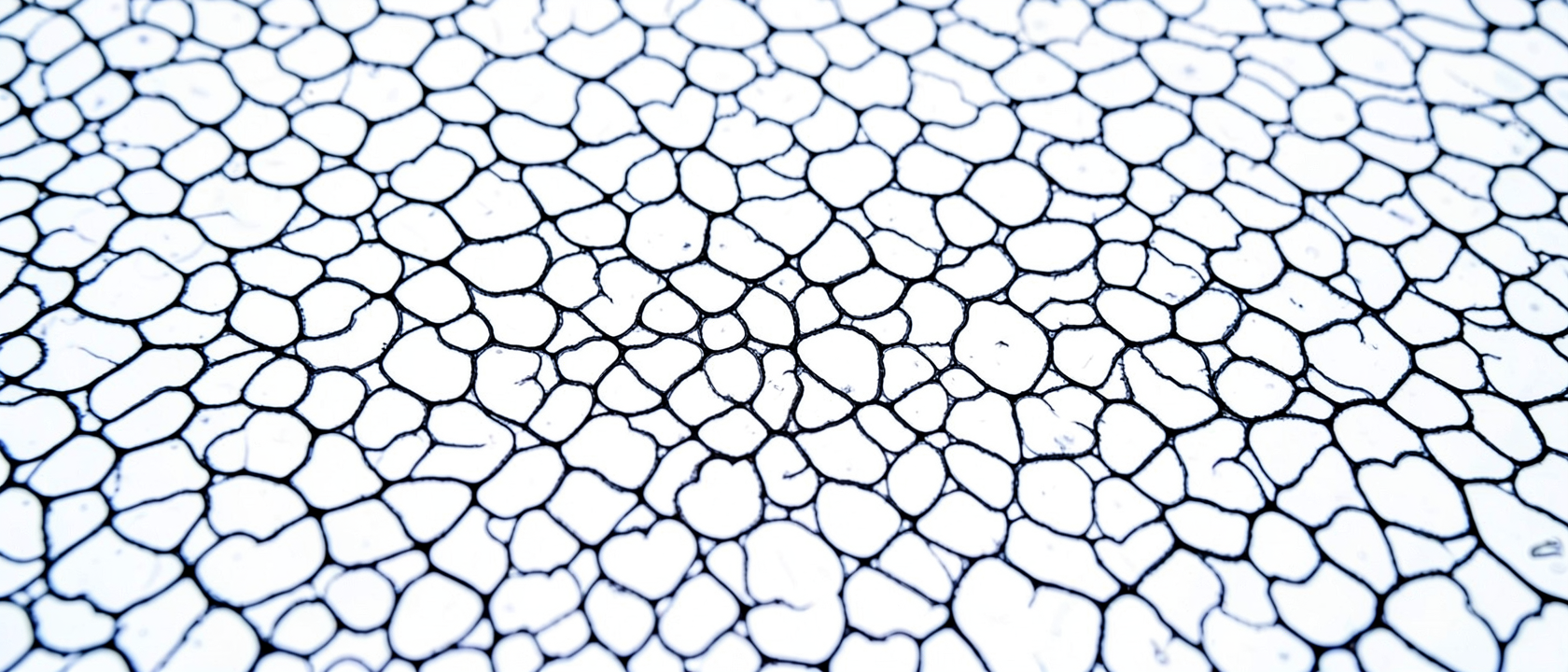
The Pattern Inside Every Breath
Show most people a photograph covered in intricately interwoven black heart shapes on a white background and they will reach for the obvious explanation: a textile print, maybe, or a piece of printmaking from an art school portfolio. Almost no one guesses what it actually is, the inner surface of a human lung, captured under a microscope.
The image came out of a lab at the University of Kansas, where assistant professor of chemical and petroleum engineering Prajna Dhar and her team, including master’s student Ashleigh Steckly and senior Ming Li Tan, were investigating something else entirely. The photograph was arresting enough to win first place in the Biophysical Society’s Art of Science Image Contest, with the winner announced on February 5, 2013, at the society’s annual conference in Philadelphia. Nobody in Dhar’s lab was trying to make art.
What You Are Actually Looking At
The substance in the image is lung surfactant: a thin molecular film that lines the walls of the alveoli, the tiny air sacs deep in the lungs where oxygen passes into the bloodstream. Surfactant is roughly 80 to 90 percent phospholipids by weight, with the remainder split between four specific proteins and neutral lipids such as cholesterol, each of those last two fractions coming in at around 10 percent of the total. The dominant molecule is one called dipalmitoylphosphatidylcholine, mercifully abbreviated to DPPC, which accounts for somewhere between 35 and 50 percent of those phospholipids depending on the individual and the measurement method.
Like dish soap, DPPC has a split personality: one end of the molecule is attracted to water, the other repels it. At an air-liquid boundary, which is exactly what the inside of an alveolus is, those molecules line themselves up into a film without any external instruction. Water-loving ends face down into the fluid, water-repelling ends face the air. Physics finds the lowest-energy solution, and that solution has a structure.
Breathing Makes the Pattern
The geometry visible in the photograph is a direct product of exhalation. As the lungs compress, the surfactant layer is squeezed and its molecules reorganize into denser, more ordered structures. Dhar described this plainly: the compression of the lung during the breathing cycle is what produces the visible pattern. The team applied a fluorescent dye that binds differently to the distinct physical phases of the surfactant film, making those phase boundaries glow under the microscope. What looks like a printed design is a frozen frame of molecular reorganization caught mid-breath.
Load-Bearing Film
Surfactant does a specific mechanical job, and the stakes become clear quickly without it. The fluid lining the alveoli generates surface tension strong enough to pull the walls of each air sac together after exhalation, the way the pages of a waterlogged book stick together when they dry. Surfactant prevents that by dynamically lowering surface tension as the lung compresses, keeping the alveoli from collapsing so they can refill with the next breath.
Premature infants, whose lungs have not yet started producing enough surfactant, develop Neonatal Respiratory Distress Syndrome for exactly this reason. The condition has historically been a significant cause of morbidity and mortality in premature infants. Synthetic surfactant treatments changed that outcome, which clarifies how essential this film actually is.
An Image from a Different Question
Dhar’s lab was not in the business of creating striking photographs. The research aimed at something practical: understanding how inhaled nanoparticles interact with lung surfactant before reaching the underlying tissue. The lungs are a likely entry point for nanoparticles into the body, and research has found that surfactant can reduce nanoparticle uptake by alveolar cells by up to two orders of magnitude, a significant finding for both pollution exposure and drug delivery. The surfactant image came out of that work as a side effect of rigorous technique applied to a different problem entirely.
What the Image Cannot Tell Us
Whether that pattern looks the same in every lung is genuinely unknown. Researchers do not yet have a clear picture of whether age, smoking history, respiratory disease, or long-term altitude exposure changes the geometry of the surfactant layer across individuals. What Dhar’s team captured is one set of lungs, one compression, one moment. The image is striking for what it reveals about a process happening billions of times in every human body, and honest about how much about that process is still being worked out.